 active
version
active
version[Mark Liberman, personal notes and links on brain anatomy and physiology, for 1998 IRCS Brain/Language reading group].
T1 is the spin lattice relaxation time for the Z component of magnetization (longitudinal magnitization).
T2 is the spin-spin relaxation time for the XY component of magnetization (transverse magnitization).
Imaging can be based on T1, T2, or spin density (rho). Sometimes apparently called "PD" because rho is not available as an ascii character. "Instrumental variables" include "TR" (repetition time), "TE" (echo time), "TI" (invesion time), "theta" (rotation angle) and T2 (because it contains a component dependent on the homogeneity of the magnetic field).
"Echo planar imaging" records an entire image in a TR period.
A magnetic resonance image is referred to as image space. Its Fourier transform is referred to as being k-space. In magnetic resonance imaging, k-space is equivalent to the space defined by the frequency and phase encoding directions. Conventional imaging sequences record one line of k-space each phase encoding step. Since one phase encoding step occurs each TR seconds the time required to produce an image is determined by the product of TR and the number of phase encoding steps. Echo planar imaging measures all lines of k-space in a single TR period.
In order to reduce imaging times, various improvements have been made in MR equipment particularly in increased magnetic gradient strength and faster rates of gradient rise. Gradient strength is measured in milli Tesla per meter (mT/m) and standard systems are around 10mT/m, whereas the new systems use gradient strengths of 20-25mT/m. The rate of rise of gradient strength or slew rate has been increased from 15-20mT/m/msec to 50-150mT/m/msec. Even further time savings are made by what is called "under the ramp" sampling. Normally, signal is acquired only whilst the gradient strength is in the static part of the pulse but with "under the ramp" sampling signal is also acquired whilst the gradient strength is rising and falling. These measures have shortened the time taken for sampling a line by a factor of 2-3. The higher gradient strengths may mean sampling at very high frequencies so that modified signal receivers are required.
In GRE imaging, a slice selective alpha-pulse takes 1msec and so a 100 line image could not be acquired in less than 150msec. Echo-planar imaging overcomes this problem by reducing the number of alpha-pulses to one. Unfortunately, the lines in this way are read under a decaying T2* curve and in tissues with a high T2*, such as muscle and liver tissue, there is virtually no signal left at the end of an echo-planar sequence so that high resolution imaging is not possible in this way. The problem is solved by introducing an additional 4-7 alpha-pulses into the sequence to make it a GRE echo-planar hybrid. This incurs very little extra time but allows the signal to be spread more evenly among the imaging lines. Such sequences are achievable on a standard scanner.
A particular application of MRI which indirectly provides information regarding relative levels of neural activity within the brain. In its most common implementation, fMRI measures a blood oxygenation level dependent (BOLD) (Kwong et al, 1992) signal. This signal reflects the relative concentration of hemoglobin not bound to oxygen (deoxy-hemoglobin), in turn reflecting blood flow and thus neural activity.
page on the ear. Another auditory system page.
The VIIIth cranial nerve exits the temporal bone by the internal auditory meatus and enters teh brainstem where the medulla meets the pons. In the brainstem, most auditory nerve fibers cross to the contralateral pathway, and travel to the midbrain and then to the temporal lobe.
Basic brainstem and midbrain way-stations are the cochlear nuclei (CN), the superior olivary complex (SOC), the nuclei of the lateral lemniscus (NLL), the inferior colliculus (IC), and the the medial geniculate body (MGB) of the thalamus.
Within the SOC, the LSO (lateral superior olive), the MSO (medial superior olive) and the MTB (medial nucleus of the trapezoid body) each has its own separate tonotopic organization (in the cat?).
IC is said to be involved in sound localization. MGB has complex responses; cells show "a notable diversity of reponse patterns (e.g., excitation, inhibition, OFF-effects, and rather complex combinations of these) [...] It is thus quite possible that many of the response patterns in the MGB are related to local inhibitory processes..." (Audition, p 293). "...cells have been found in the MGB that respond to complex sound patterns (such as vocalization of other behaviorally significant sounds), whereas they do not respond to pure tones."
For all mammls the auditory cortex is in a subsilvian position. In the carnivores, it is largely spread over the convex surface of temporal regions; in primates, it is more or less buried in the sylvian fissure. Because of the coexisting direct and crossed connections in the auditory pathways, each cochlea is apparently universally represented bilaterally in the auditory cortex. (Audition, p. 296)
The primary auditory cortex contains several distinct tonotopic representations (what and where??). Layer VI projects back to the medial geniculate nucleus and layer V projects to the inferior colliculus. Primary auditory cortex = Brodmann's areas 41 and 42 (superior temporal gyrus) (Transverse temporal gyrus or Heschel's gyrus). Area 41: contains the central "Koniocortex" and is buried in the inferior operculum of the lateral fissure. Tonotopic organization of primary auditory cortex: (data based on PET scans): There is a medial to lateral organization: Low frequency tones are represented laterally and high frequency tones are represented medially.
Relevant pathologies: verbal and non-verbal auditory agnosia.
The cortex is a sheet of neurons, about 2.5 sq. ft in area, 2-4 mm thick, organized into a series of laminar and vertical (columnar) features. The structure varies from region to region
Neocortex: 90% of cortical area Paleocortex: some restricted parts on base of telencephalon: mostly olfactory cortex. Archicortex: hippocampus (associated with memory and the limbic system.
Types of cortical neurons: 1. Pyramidal cells: main output neuron; some can be very large up to 100 µm in dia.; i.e., giant Betz cells of motor cortex. 2. Stellate cells (or granule cells) main interneurons of cortex (local circuit neurones). Small (less than 10 µm), multipolar with short axons that do not leave the cortex.
The different layers have different cell types and different input-output characteristics. Neocortex has six layers. Hippocampal cortex (Archicortex) has 3 layers, referred to as allocortex. Olfactory cortex (paleocortex) has variable number of layers
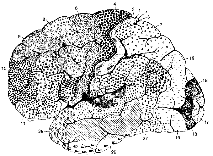
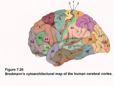
The neocortex is heterogeneous and can be divided into various regions based on cytoarchitectonic features (cell size, cell density, cell type etc.). Six layers are not easily seen everywhere. Brodmann (1909) identified 50 (52?) different regions.
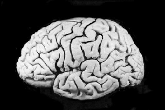
Gyri: the folds or convolutions of the cerebral cortex Sulci: the intervening groves between the gyri on the surface of the cerebral hemisphere Fissure: a cleft that separates large components of the brain (not disjoint from sulcus).
Major sulci and fissures
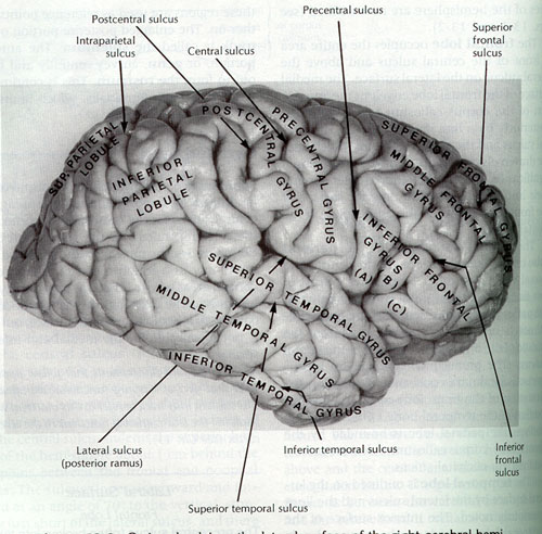
Lobes of the Cerebral Hemispheres
Gyri and Sulci of the Cerebral Hemispheres
Lateral surface
Frontal Lobe
- Precentral sulcus:- runs anterior and parallel to the central sulcus
- Precentral gyrus:- between the central and precentral sulci
- Superior, Middle and Inferior Frontal gyri:- divided by the superior and inferior sulci. The inferior frontal gyrus is divided into opercular, triangular and orbital portions by the anterior and ascending rami of the lateral sulcus.
Parietal Lobe
- Postcentral sulcus:- runs posterior and parallel to the the central sulcus
- Postcentral gyrus:- bounded by the central and postcentral sulci
- Intraparietal sulcus:- runs posteriorly from the postcentral sulcus and divides the parietal lobe (excluding the postcentral gyrus) into superior and inferior parietal lobules. The inferior parietal lobule is divided into the "supramarginal gyrus," the "angular gyrus," and an additional posterior convolution
Temporal Lobe
- Superior and Inferior Temporal sulci:- divide the lateral surface of the temporal lobe into
- Superior, Middle and Inferior temporal gyri.
- Insula The insula is concealed by the frontal, parietal and temporal opercular on the lateral surface. It is outlined by a circular sulcus and is divided into two regions by a central sulcus. Short gyri lie in front of the central sulcus and long gyri lie behind the sulcus.
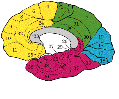
Medial and Inferior Surfaces

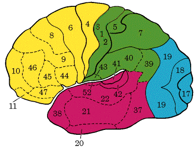
Primary visual cortex -- BA 17, located on the walls of, and surrounding the calcarine sulcus. - also known as the striate area (because the fourth layer of cells, involving projections from the thalamus, is so dense that it can be seen even without a microscope). Visual Association Cortex:- this area is found at Brodmann areas 18 and 19, surrounding the visual area on the medial and lateral surfaces of the hemisphere, receives fibres from the visual area as well as other cortical areas and the pulvinar of the thalamus.Other secondary visual areas: 20, 21, 37?
Frontal Eye Field:- the lower part of BA 8 in the middle frontal gyrus, extending into the superior frontal gyrus.
Prefrontal Cortex:- BA 9,10,11 and 12, including most parts of the superior, middle and inferior frontal gyri, the orbital gyri, the medial frontal gyrus and the anterior half of the cingulate gyrus.
Primary auditory cortex -- BA 41 and 42, mostly located in the ventral wall of the lateral sulcus.(Is 42 primary or secondary auditory cortex?) It also includes the transverse temporal gyri of Heschl, buried in the wall of the lateral fissure. Auditory Association Cortex: BA 22, on the floor of the lateral sulcus and the lateral surface of the superior temporal gyrus. Wernicke's area: (part of?) BA 22 in the (posterior part of the) superior temporal gyrus, and 39 and 40 in the supramarginal & angular gyri.
Primary Motor Area:- BA 4 in the precentral gyrus, the anterior wall of the central sulcus and the anterior part of the superior paracentral lobule. Second Motor Area:- area ventral to the central sulcus in the dorsal wall of the lateral sulcus. Supplementary Motor Area:- part of BA 6 and 8 on the medial part of the superior frontal gyrus, in front of the paracentral lobule (8 is eye movement?) Premotor Area:- BA 6 in the anterior part of the precentral gyrus and the posterior part of the superior frontal gyrus. Broca's area -- 44 -- 45, in the opercular and triangular portions of the inferior frontal gyrus. Tertiary motor areas: 9,10, 11, 45, 46, (?). According to Luria, 1973, premotor cortex is responsible for the conversion of individual motor impulses into consecutive kinetic melodies.
Primary somatosensory area -- 1,2,3. Second Somesthetic area:- a small area on the dorsal wall of the lateral sulcus - the parts of the body are represented bilaterally, but contralateral representation is dominant. Primarily involved in the less discriminative aspects of sensation. Somesthetic Association Cortex:- this area mostly corresponds to Brodmann areas 5 and 7 and is mainly located in the superior parietal lobule and the precuneus.
Taste Area: BA 43, in the inferior part of the parietal lobe, posterior to the general sensory area for the mouth.
Q: what BA is PT ?
Q: two pictures show BA 44 posterior to 45, one shows it superior (and even a bit anterior). Who's right? or does it vary?
Adjacent to primary auditory cortex (area 41), inside sylvian fissure, at back of temporal lobe. Generally larger on the left side. Planum Parietale (extension of the same structure? into parietal lobe) is generally larger on the right side.
According to Calvin/Ojemann, asymmetry is shared by chimps and orangutans but not gorillas.
Some reports of lack or reversal of PT asymmetry in dyslexics and in schizophrenics (another study).PT is said to be enlarged in Williams Syndrome.
However, a recent study (Asymmetries in dyslexics) reports about 70-80% of both dyslexics and controls with PT L>R, and about 50-60% of both groups with PP R>L.
A 1995 study based on size claims that PT asymmetry is greater in musicians than in nonmusicians, and greatest of all in those with perfect pitch. A 1998 functional study showed that PT lights up in perfect pitchers NOT in perception but only in naming of pitches.
What Brodmann area is PT (and PP)?
Coleman, John. "Cognitive Reality and the Phonological Lexicon: A Review." J. Neurolinguistics 11.3, pp. 295-320, 1998.
Coleman suggests that "entires in the lexicon are very probably auditory, and improbably articulatory." As part of his argument, he suggests that "there is strong evidence for only a single phonological lexicon in speech -- albeit one which is duplicated bilaterally -- in the superior temporal gyri."
This does not seem to be consistent with Zatorre et al. 1996, who describe a PET study comparing lexical decision (to spoken words) with passive listening, which yielded:
| Region | Brodmann Area | x | y | z | t value |
| (Blood flow increases) | |||||
| 1. M Posterior cingulate gyrus | 23 | 3 | -28 | 22 | 4.28 |
| 2. M Anterior cingulate gyrus | 24 | 3 | 3 | 24 | 4.07 |
| 3. R Middle frontal gyrus | 9 | 29 | 5 | 35 | 3.91 |
| (Blood flow decreases) | |||||
| 4. R Insula | - | 38 | 6 | 8 | 4.27 |
| 5. L Insula | - | -34 | -21 | 21 | 4.06 |
| 6. R pre/post central gyrus | 4/3 | 56 | -13 | 44 | 3.91 |
| 7. L Middle frontal gyrus | 6 | -35 | 5 | 53 | 3.80 |
| 8. R Transverse temporal gyrus | 41 | 44 | -21 | 13 | 3.70 |
I take it that "superior temporal gyrus" would be BA 22, which is not active in this comparison. It would be better to have a nonspeech or at least nonlexical comparison, but shouldn't the "phonological lexicon" be more active in a lexical decision task than in passive listening? I guess maybe not, if the access is unproblematic and entirely automatized, but still...
It may also worth observing that the anterior cingulate gyrus underlies Broca's area (?)In other tasks, this studies identifies Broca's area in terms of coordinates (-48 3 24) (-43 5 27) (-56 6 29). What is in between? The supplementary motor area and the premotor cortex seem to meet just about "outside" the cingulate gyrus (?)
Other cingulate gyrus stuff: "...you seem to use some regions of the midline regions of the frontal lobe, the cingulate gyrus just in front of the corpus callosum, to stay alert for a class of words -- such as when you are looking for Italian surnames while scanning a telephone book." (CWNB p. 82). So maybe it's just attentional? But "The brain area immediately below the supplementary motor area, the cingulate gyrus, is part of the 'emotional' limbic system." (CWNB p. 229). And "...the cingulate gyrus has shown activity with word reading in blood flow studies with normal volunteers." CWNB p. 228). "Monkey neurons in that region [the supplementary motor area and the cingulate gyrus] change their activity when the monkey hears vocalizations -- often selectively, responding only to vocalizations of their own species." (CWNB p. 228).
The cingulate gyrus is an arched convolution that lies next to the corpus callosum and is separated from it by the sulcus of the corpus callosum. It begins below the rostrum, curves around in front of the genu, extends along the trunk, and then turns downward behind the splenium of the corpus callosum, where it is connected by the narrow "isthmus of the cingulate gyrus" to the parahippocampal gyrus. It is separated from the "medial frontal gyrus" by the cingulate sulcus and from the "precuneus" by the more variable "suprasplenia sulcus." It has many connections with the anterior thalamic nuclei and is often considered to be part of the "limbic system."
In favor of the attentional interpretation -- anterior cingulate gyrus is active during both color and spatial stroop tasks, and also in dual/alternating English/Spanish naming.
The Anterior Cingulate Gyrus is part of the lymbic sytem which mediates emotions and aspects of learning and memory. It is associated with vigilance and is active in almost any task that involves an increase in concentration and/or attention.
Zatorre et al. conclude that "neural processes in the superior temporal gyri are initially responsible for perceptual analysis of the complex incoming speech stream. [more].
Zatorre, Robert J., E. Meyer, A. Gjedde, and A.C. Evans. "PET Studies of Phonetic Processing of Speech: Review, Replication, and Reanalysis." Cerebral Cortex 6.1, pp. 21-30, 1996.
lies at the boundary between the occipital, temporal, and postcentral regions of the hemisphere, where the cortical areas for visual, auditory, vestibular, cutaneous, and proprioceptive sensations overlap. Supramodal in function: plays a special role in inter-analyzer syntheses. The angular gyrus, as part of the inferior parietal lobule, is the association area of association areas and allows cross modal transfer and associations between either vision or touch and hearing. Important in the processing of associating a heard name to a seen or felt object, and probably also important for associations in the reverse direction.
Ojemann found that "patients whose sites for reading were in the superior temporal gyrus, with naming in the middle temporal gyrus, had high verbal IQs. And he found the reverse in patients with low verbal IQs." (Conversations with Neil's Brain, p. 236).
functional anatomy of auditory word processing
imaging of auditory hallucinations